First-in-human, before the first patient.

ERIDAN tests coronary devices on fresh-frozen human hearts and reads the result with micro-CT, intravascular imaging and histology. An Anatomy & Imaging Hub in Torino, founded by interventional cardiologists.

Fig. 01 · Micro-CT reconstruction of a calcified coronary segment, rebuilt slice by slice. Illustration.

From clinical intuition to industrial evidence.

A device idea usually reaches the bench late and the patient early. ERIDAN puts a human coronary artery in between. The same team designs the study, runs it on the ex vivo bench, images the result and returns data an engineer can use.
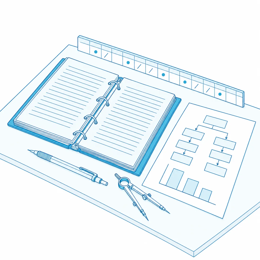
Study design: an open protocol binder with a flowchart of study arms, a small bar chart and drafting dividers on a desk
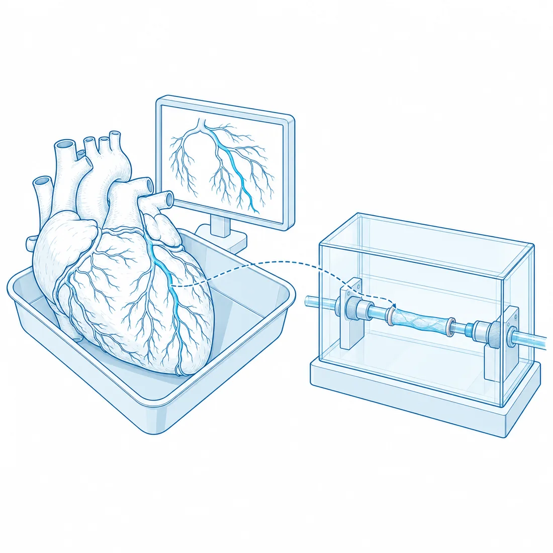
Ex vivo bench: a human heart on a tray beside a CT angiography display, and the isolated coronary segment being mounted in the perfusion chamber
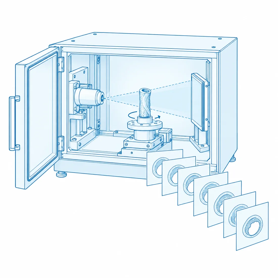
Micro-CT: a scanner cabinet open on the rotating stage holding the segment between X-ray source and detector, a stack of slices emerging
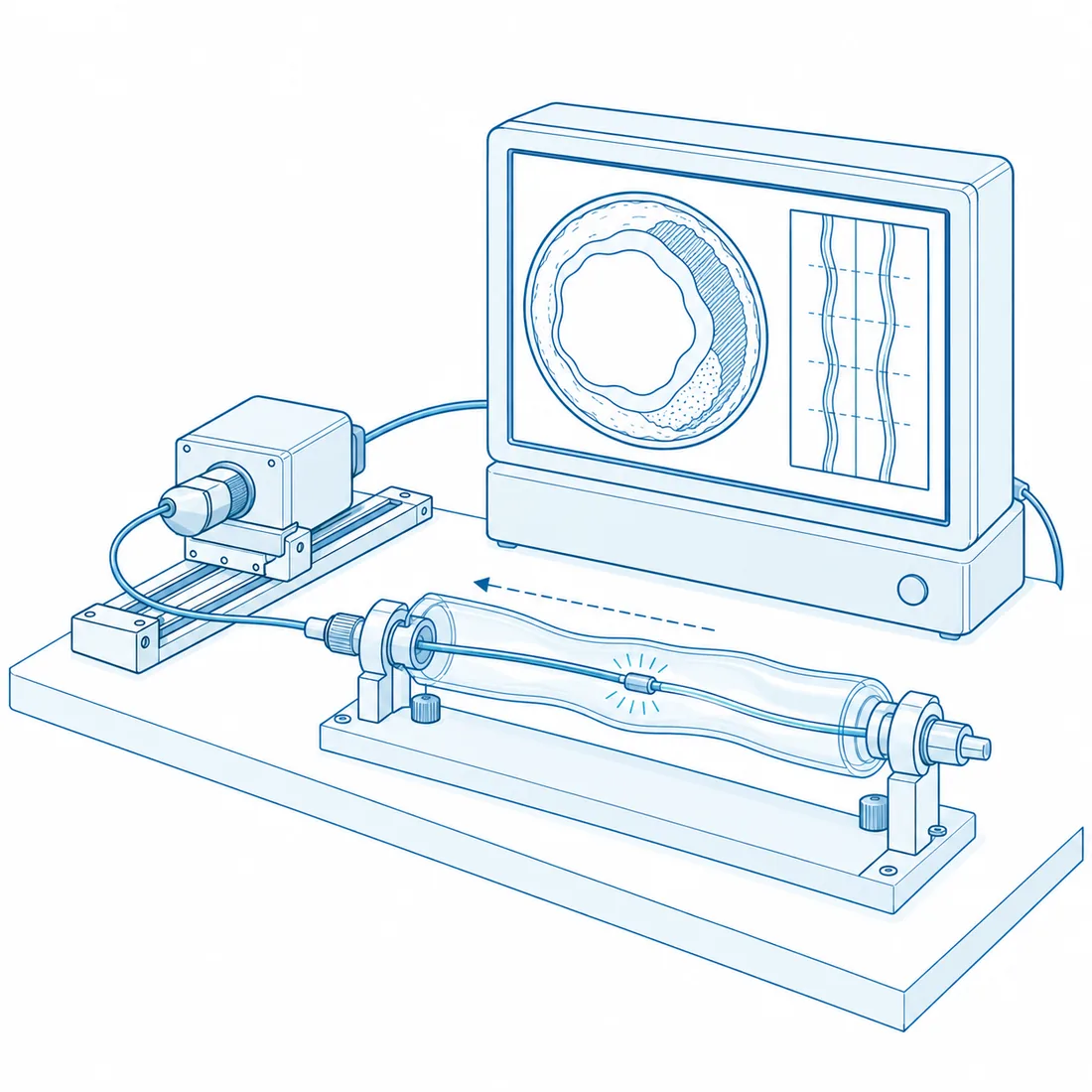
Intravascular imaging: an OCT catheter inside the vessel segment with a pullback unit and a console showing the cross-section frame
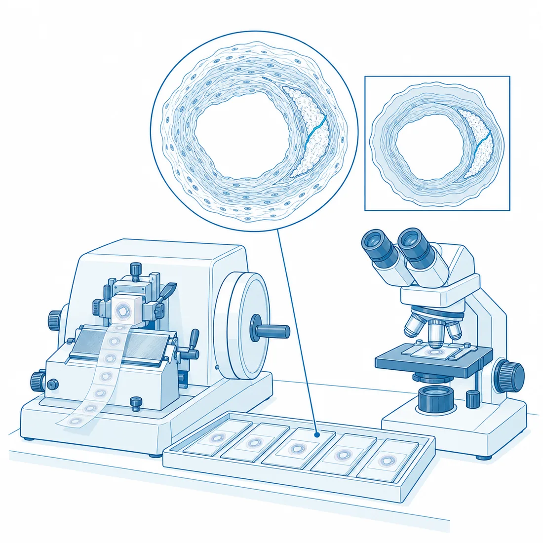
Histology: a microtome cutting a paraffin block, slides in a tray, a microscope and one magnified slide showing the vessel layers
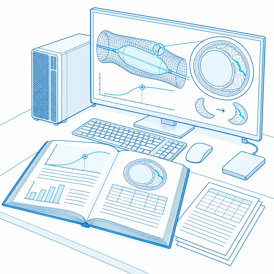
Data, models, evidence: a bound report with charts beside a laptop showing a wireframe digital twin of the vessel and a dose–response curve
Fig. 02 · Study design
  1. Study design

    Protocol, endpoints, sample size and the ethics dossier, written by the clinicians who will run the bench. Kick-off with your R&D team, signed protocol as the output.

  2. Ex vivo bench

    Fresh-frozen human hearts selected by CT angiography. Coronary segments with native calcium, real bifurcations, real tortuosity. Devices deployed as in the cath lab, on a bench that reproduces pressure and flow.

  3. Micro-CT

    30 µm voxels on the treated segment, before and after: fracture mapping, strut apposition, calcium morphology. Mechanical tests under continuous scanning when the question is dynamic.

  4. Intravascular imaging

    OCT and IVUS pullbacks read mm by mm: lumen, external elastic membrane, struts, calcium arc and thickness, fused with the micro-CT volume.

  5. Histology

    Sections read by a university hospital pathology unit and matched frame by frame to the imaging.

  6. Data, models, evidence

    A publication-ready report with raw data and video, and a device digital twin trained on the measurements, so the next design and the next technique start from numbers.

From the cath lab to the bench.

Every field is run by the same people who perform PCI every week. Pick a field: the plate changes and the list of what we do in it opens.
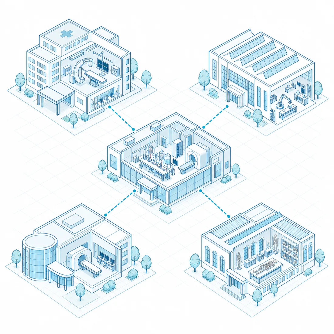
Isometric map of the ERIDAN hub: a laboratory with a perfusion bench and a micro-CT at the centre, linked to a university hospital, an engineering hall, an imaging centre and an anatomy institute
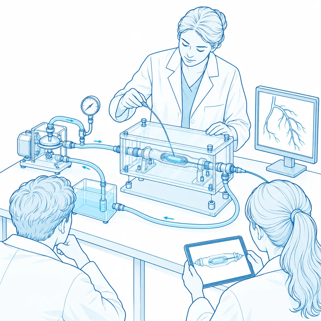
A teaching session: an instructor at the ex vivo bench guiding a catheter into the perfusion chamber while two students watch, one with a tablet, a monitor showing a coronary tree
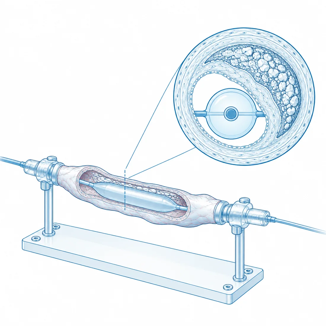
An isolated coronary segment on a mount with a balloon catheter inside, beside a large cross-section showing an eccentric calcium crescent
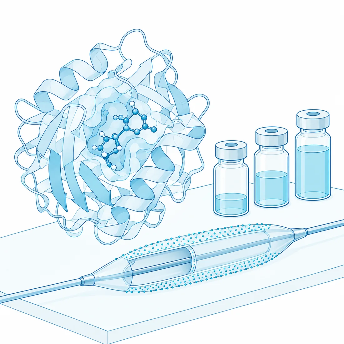
A small molecule in a protein pocket beside a drug-coated balloon in cutaway and three vials
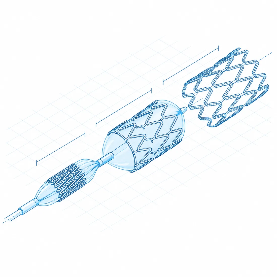
Exploded view of a coronary stent: crimped, expanded and as a finite-element wireframe, with dimension lines
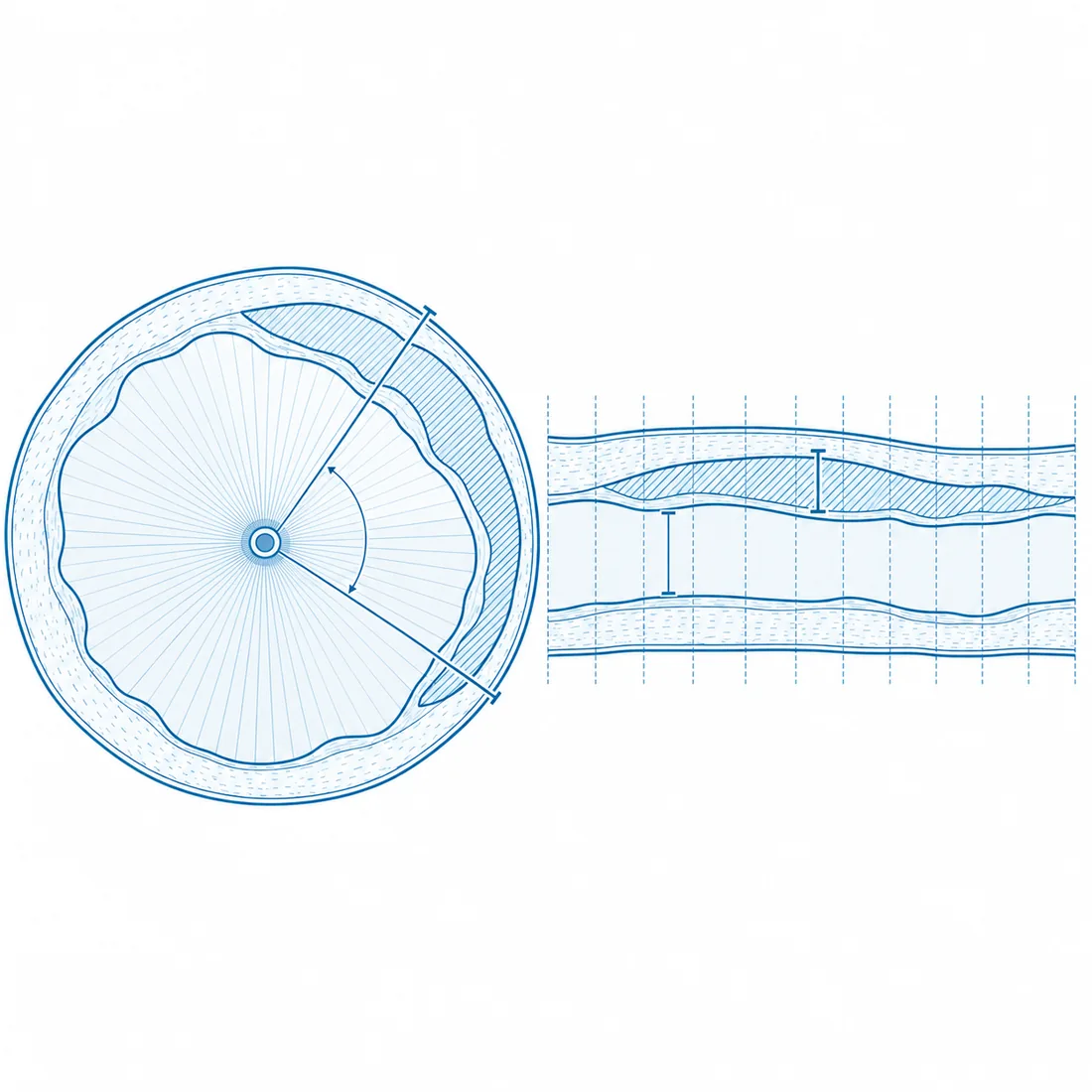
Quantitative imaging plate: an OCT frame with lumen and vessel contours and a longitudinal pullback strip with calipers
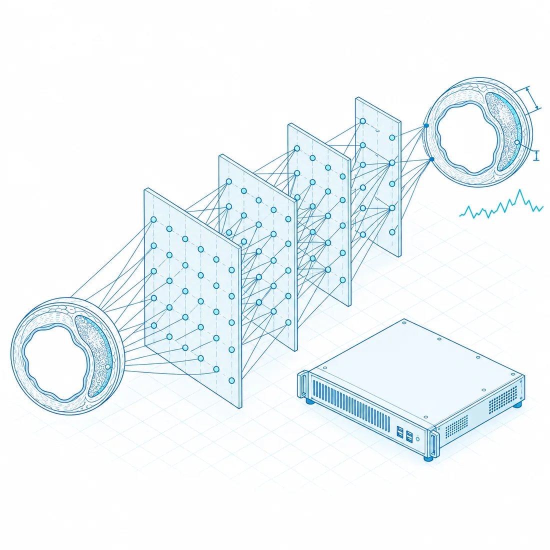
A neural network drawn as stacked translucent layers of nodes, taking an OCT-like vessel cross-section as input and returning contours and measurements, beside a small compute node
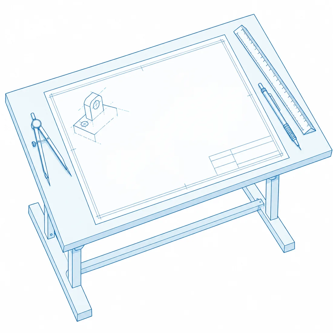
A drafting table with a blank technical sheet, dividers, a scale rule and an unfinished device sketch
Fig. 03 · Hub · Illustration

Four ways to work with us.

Every engagement is modular. A start-up with a first prototype and a multinational strengthening a CE or FDA dossier buy different pieces of the same bench.
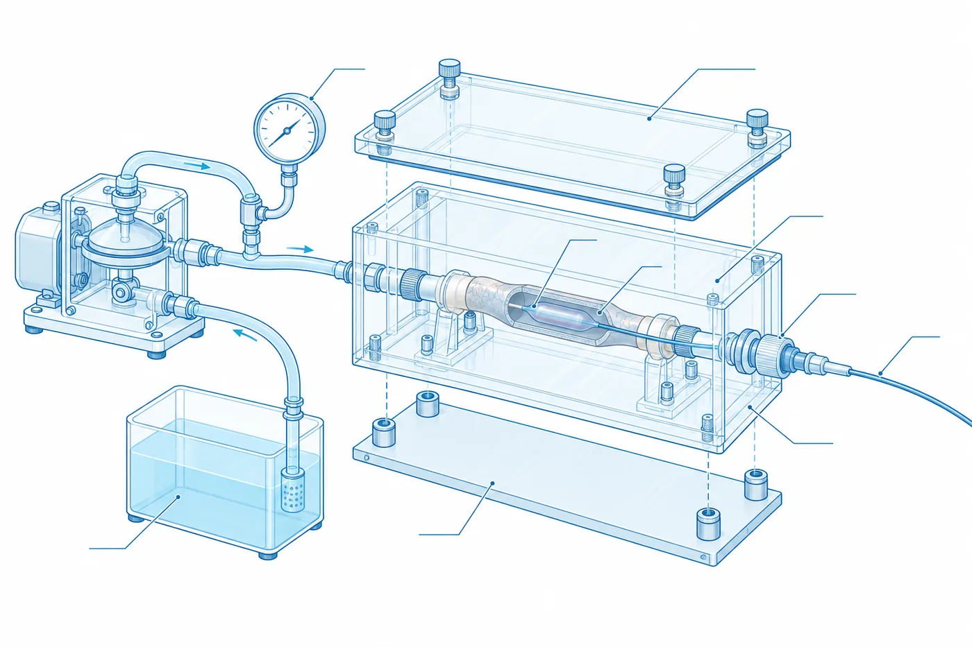
Exploded drawing of the ex vivo bench: pulsatile pump and reservoir, pressure gauge, a transparent perfusion chamber holding a coronary segment with a balloon catheter inside
Fig. 04 · The ex vivo bench: pulsatile perfusion, pressure line, coronary segment in the chamber, catheter access. Schematic.

Full preclinical study

Fresh-frozen anatomy, the complete procedure in the Cadaver Lab, OCT, IVUS and micro-CT, core-lab analysis and a publication-ready report. For CE and FDA dossiers and preclinical phases.

Predictive report

A computational model of your device in variable anatomical scenarios, built with bioengineering partners. Usually added to a full study.

Proof of concept

A half-day feasibility test for an early prototype: basic mechanics plus one imaging acquisition, before investing in a full study.

Regulatory data pack

Micro-CT images, statistics and histomorphometric cross-sections formatted as a dossier ready to attach to a CE or FDA submission.

And beyond testing

Product launch events and master courses in the Academy; redesign consulting, rapid prototyping and licensing when a study shows how a device could be better; molecular design when the question is the drug on the device.

How a study runs

  1. Scientific kick-off: endpoints, device, anatomical scenarios, signed protocol
  2. Anatomical procurement: fresh-frozen preparation, cold chain, Cadaver Lab preparation
  3. Experimental set-up: perfusion system, OCT and IVUS calibration, micro-CT geometry, baseline pilot
  4. Experiment: your device, imaging before, during and after, full video; remote participation possible
  5. Micro-CT and histomorphometry: high-resolution scan, 3D cross-sections, histology if requested
  6. Core-lab analysis: dissection, expansion, plaque modification, release uniformity, statistics
  7. Delivery and debrief: report, raw data and video; one hour with your R&D team

FRACTAL

FRACTure Analysis after Lithotripsy

Ex vivo study · protocol stage · 2026

Lithotripsy fractures calcium, and the fracture pattern decides how the vessel yields. FRACTAL maps that pattern on micro-CT, fracture by fracture.
Design
Ex vivo, two arms: intravascular lithotripsy versus a non-compliant balloon, on calcified human coronary segments from fresh-frozen hearts.
Primary readout
Circumferential fracture map from micro-CT: number, depth and angular position of calcium fractures, with dose–response across pulse counts.
Secondary readouts
OCT and IVUS lumen gain and fracture visibility; histology of the treated segment.
What the partner receives
A fracture atlas per lesion, the dose–response curve, and a device digital twin trained on the measurements.
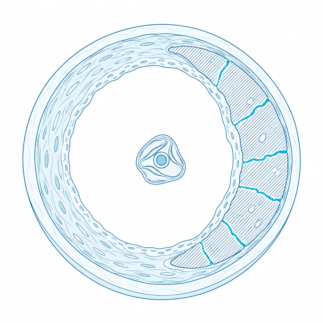
Schematic cross-section of a calcified coronary artery after lithotripsy: an eccentric crescent of calcium crossed by radial fractures, a deflated balloon in the lumen
Fig. 05 · Calcified segment after intravascular lithotripsy, cross-section. Schematic drawing, not study data.

Device digital twin

Schematic twin of a lithotripsy balloon in a calcified segment: inflation, pressure pulses, fracture propagation and post-dilatation, with a magnified view of the contact between membrane and calcium. The sequence runs on its own. Geometry, forces and fracture count are schematic; study data will replace them.

Pressure
0.0 atm
Pulses
00 / 10
Fractures
0
Lumen gain
+0%

Fig. 06 · Device digital twin, schematic simulation. Study data will replace geometry, forces and fracture count.
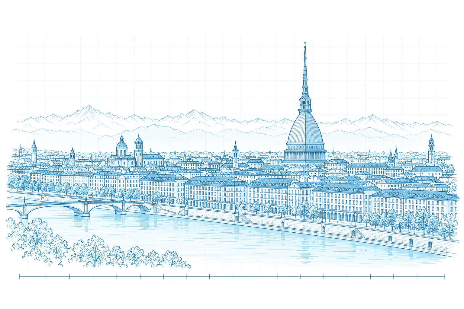
Line drawing of Turin seen from the Po: the Mole Antonelliana, the riverside buildings and the Alps behind the city
Fig. 07 · Turin from the Po. Illustration.

Torino, 45°04′N.

Two hours from Milano, three from Lyon, four from Zürich. A university hospital with a cath lab and a pathology unit, a molecular imaging centre with a micro-CT, a polytechnic with a bioengineering department, and an additive-manufacturing competence centre, all within the same city.

Micro-CT
30 µm voxels at a university molecular imaging facility
Histology
University hospital pathology unit
Cath labs
Interventional cardiology units of the Torino area
Engineering
Bioengineering and additive manufacturing partners
Fig. 08 · The Torino network around the hub.

Start a study.

Tell us the device or the molecule and the question you need answered. We reply with a first design and a realistic timeline.

The form opens your e-mail client with the message prefilled. Nothing is sent from this page.